

3B Scientific Hand Skeleton Model with Ligaments and Muscles

Grades 6–12 Study the human hand in great detail with this anatomically correct model. The bones, muscles, tendons, ligaments, nerves, arteries, and veins are all displayed in this four-part model of the hand and lower forearm.
The dorsal side of the hand shows the extensor muscles as well as portions of the tendons at the wrist as they pass under the extensor retinaculum. The palmar face of the hand is represented in three layers. The first two are removable so the deeper anatomical layer can be viewed so student scan study the intrinsic muscles and deep palmar arterial arch in addition to other important structures.
This high-quality model includes free access to the anatomy course 3B Smart Anatomy, which includes 23 digital anatomy lectures, 117 different virtual anatomy models, and 39 anatomy quizzes to test student knowledge using the Complete Anatomy app by 3D4Medical. Allow extra delivery time.


| Brand | American 3B Scientific |
|---|---|
| Item Weight | 2 lbs |
| Length | 14.00 |
| Width | 7.00 |
| Height | 9.00 |
| Age | 11 yrs-18 yrs |
| Grade | 6-12 |
| Prop 65 |
|
| School Type | High School, Middle School |

-
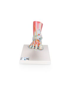 3B Scientific Foot Skeleton Model with Ligaments and Muscles $406.98Product Number: NE40266In Stock